
Herpes hipertrófico nasal em paciente vivendo com HIV/AIDS
DOI:
https://doi.org/10.5935/2764-734X.e20230928Palavras-chave:
Infecções oportunistas relacionadas com a AIDS, Dermatopatias virais, Infecções por herpesvírus, Medicamentos antivirais, Imiquimode, Procedimentos de cirurgia plástica, Relato de casoResumo
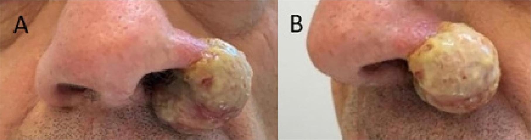
Lesões hipertróficas causadas pelo vírus herpes simplex são apresentações incomuns descritas usualmente em pacientes imunossuprimidos. Estas condições são frequentemente desafiadoras, tanto no seu diagnóstico quanto no manejo. Falhas terapêuticas aos antivirais de primeira linha são comuns e outras estratégias de tratamento são frequentemente necessárias, como o uso de imunomoduladores e até a ressecção cirúrgica. Aqui descrevemos o caso de uma pessoa vivendo com HIV que foi diagnosticada com herpes hipertrófico em nariz, refratário aos tratamentos convencionais.
Downloads
Referências
1. Beauman JG. Genital herpes: a review. Am Fam Physician. 2005;72(8):1527-34.
2. Sasso BM, Florence ME, Magalhaes RF, Velho PE, de Souza EM, Cintra ML, et al. Herpes simplex virus mucocutaneous tumoural lesions–systematic review. J Clin Virol. 2020;123:104246. Available at: https://www.sciencedirect.com/science/article/abs/pii/S1386653219302768?via%3Dihub
3. Barde C, Piguet V, Pechère M, Masouyé I, Saurat JH, Wunderli W, et al. Management of resistant mucocutaneous herpes simplex infections in AIDS patients: a clinical and virological challenge. HIV Med. 2011;12(6):367-73.
4. Wald A, Corey L. Persistence in the population: epidemiology, transmission. In: Arvin A, Campadelli-Fiume G, Mocarski E, Moore PS, Roizman B, Whitley R, Yamanishi K, editores. Human Herpesviruses: Biology, Therapy and Immunoprophylaxis. Cambridge University Press; 2007. Chapter 36. Available at: https://www.ncbi.nlm.nih.gov/books/NBK47447/
5. Severson JL, Tyring SK. Relation between herpes simplex viruses and human immunodeficiency virus infections. Arch Dermatol. 1999;135(11):1393-7.
6. Chiu CY, Randhawa G, Nada K, Tomczak E, Feinstein A, Hennessey K. A nasal hypertrophic lesion as a presentation of herpes simplex virus. IDCases. 2019;15:e00512. Available at: https://www.sciencedirect.com/science/article/pii/S2214250919300162?via%3Dihub.
7. Strehl JD, Mehlhorn G, Koch MC, Harrer EG, Harrer T, Beckmann MW, et al. HIV-associated hypertrophic herpes simplex genitalis with concomitant early invasive squamous cell carcinoma mimicking advanced genital cancer: case report and literature review. Int J Gynecol Pathol. 2012;31(3):286-93.
8. Cury K, Valin N, Gozlan J, Jacquier I, Boutolleau D, Guegan S, et al. Bipolar Hypertrophic Herpes: An Unusual Presentation of Acyclovir-Resistant Herpes Simplex Type 2 in a HIV-Infected Patient. Sex Transm Dis. 2010; 37(2):126-128
9. 9. Beasley KL, Cooley GE, Kao GF, Lowitt MH, Burnett JW, Aurelian L. Herpes simplex vegetans: atypical genital herpes infection in a patient with common variable immunodeficiency. J Am Acad Dermatol. 1997;37(5):860-3.
10. Deza G, Martin-Ezquerra G, Curto-Barredo L, Villar Garcia J, Pujol RM. Successful treatment of hypertrophic herpes simplex genitalis in HIV-infected patient with topical imiquimod. J Dermatol. 2015;42(12):1176-8.
11. Arshad Z, Alturkistani A, Brindley D, Lam C, Foley K, Meinert E. Tools for the diagnosis of herpes simplex virus 1/2: systematic review of studies published between 2012 and 2018. JMIR Public Health Surveill. 2019;5(2):e14216. Available at: https://publichealth.jmir.org/2019/2/e14216/
12. Smith KJ, Skelton III HG, Frissman DM, Angritt P. Verrucous lesions secondary to DNA viruses in patients infected with the human immunodeficiency virus in association with increased factor XIIIa-positive dermal dendritic cells. J Am Acad Dermatol. 1992;27(6):943-50.
13. Abbo L, Vincek V, Dickinson G, Shrestha N, Doblecki S, Haslett PA. Selective defect in plasmacyoid dendritic cell function in a patient with AIDS-associated atypical genital herpes simplex vegetans treated with imiquimod. Clin Infect Dis. 2007;44(3):e25-7.
14. Arinze F, Shaver A, Raffanti S. Surgical excision for recurrent herpes simplex virus 2 (HSV-2) anogenital infection in a patient with human immunodeficiency virus (HIV). Infection. 2017;45:705-7.
15. Shelley WB. Surgical treatment for recurrent herpes simplex. Lancet. 1978; 11;312(8098):1021-2.
16. Simonsen M, Nahas SC, Silva Filho EV, Araújo SE, Kiss DR, Nahas CS. Atypical perianal herpes simplex infection in HIV-positive patients. Clinics. 2008;63:143-6.
Downloads
Publicado
Como Citar
Edição
Seção
Licença
Copyright (c) 2023 Infectologia em Evidência

Este trabalho está licenciado sob uma licença Creative Commons Attribution 4.0 International License.
Todos os usuários podem ler, baixar, compartilhar e adaptar esta produção científica livremente para quaisquer fins (mesmo que comerciais), desde que seja dado o devido crédito aos autores e à publicação original e que qualquer alteração seja devidamente indicada.
> Ética
Todos os artigos publicados na Revista gozam de uma aprovação ética do Sistema Nacional de Ética em Pesquisa (antigo sistema CEP/CONEP) com base na Lei Federal 14.874/24 e outras regulamentações específicas brasileiras ou documento semelhante atestando a ciência e autorização por parte da instituição de origem no caso de trabalhos estrangeiros.
Os autores declaram não haver nenhum tipo de patrocínio ou conflito de interesses, salvo indicação em contrário no corpo do artigo.
Vale ressaltar que os relatos de caso são um valioso recurso de aprendizado para a comunidade científica, mas não devem ser utilizados isoladamente para guiar opções diagnósticas ou terapêuticas na prática clínica ou em políticas de saúde.


