
Coinfecção de vírus monkeypox e HIV-1 em paciente gravemente imunossuprimido: uma evolução catastrófica
DOI:
https://doi.org/10.5935/2764-734X.e20231232Palavras-chave:
Vírus da varíola dos macacos, HIV-1, Infecções oportunistas relacionadas com a aids, Relato de casoResumo
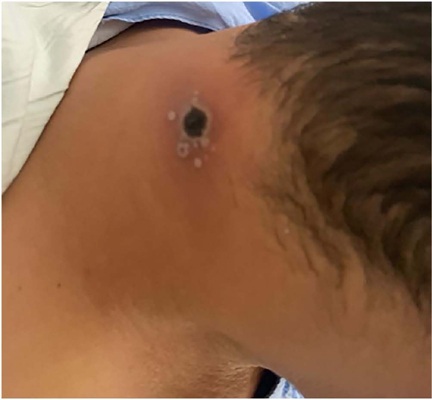
A monkeypox (MPOX) é uma doença causada pelo vírus monkeypox (MPXV), endêmico desde 1970 na África Central e Ocidental, com poucos surtos relatados fora desse continente. Em maio de 2022, observou-se mundialmente um aumento no número de casos da doença, com maior prevalência em homens que fazem sexo com homens e comprovada transmissão através de contato direto, incluindo exposição sexual. Nesse contexto, evidenciou-se a associação da MPOX com infecções sexualmente transmissíveis, tal como a infecção pelo HIV. O caso apresentado retrata a coinfecção de MPXV e HIV-1 em paciente gravemente imunossuprimido, ressaltando a importância da interação de ambas as infecções no prognóstico clínico.
Downloads
Referências
1. Petersen E, Kantele A, Koopmans M, Asogun D, Yinka-Ogunleye A, Ihekweazu C, et al. Human monkeypox: epidemiologic and clinical characteristics, diagnosis, and prevention. Infect Dis Clin North Am. 2019 Dec;33(4):1027-43. DOI: 10.1016/j.idc.2019.03.001
2. Gong Q, Wang C, Chuai X, Chiu S. Monkeypox virus: a re-emergent threat to humans. Virol Sin. 2022 Aug;37(4):477-82. DOI: 10.1016/j.virs.2022.07.006
3. Kumar N, Acharya A, Gendelman HE, Byrareddy SN. The 2022 outbreak and the pathobiology of the monkeypox virus. J Autoimmun. 2022 Jul;131:102855. DOI: 10.1016/j.jaut.2022.102855
4. Thornhill JP, Barkati S, Walmsley S, Rockstroh J, Antinori A, Harrison LB, et al. Monkeypox virus infection in humans across 16 countries - April-June 2022. N Engl J Med. 2022 Aug;387(8):679-91. DOI: 10.1056/NEJMoa2207323
5. Vusirikala A, Charles H, Balasegaram S, Macdonald N, Kumar D, Barker-Burnside C, et al. Epidemiology of early monkeypox virus transmission in sexual networks of gay and bisexual men, England, 2022. Emerg Infect Dis. 2022;28(10):2082-6. DOI: 10.3201/eid2810.220960
6. Philpott D, Hughes CM, Alroy KA, Kerins JL, Pavlick J, Asbel L, et al. Epidemiologic and clinical characteristics of monkeypox cases - United States, May 17-July 22, 2022. MMWR Morb Mortal Wkly Rep. 2022 Aug;71(32):1018-22. DOI: 10.15585/mmwr.mm7132e3
7. León-Figueroa DA, Barboza JJ, Garcia-Vasquez EA, Bonilla-Aldana DK, Diaz-Torres M, Saldaña-Cumpa HM, et al. Epidemiological situation of monkeypox transmission by possible sexual contact: a systematic review. Trop Med Infect Dis. 2022;7(10):267. DOI: 10.3390/tropicalmed7100267
8. Ministério da Saúde (BR). Boletim Epidemiológico Especial: MPOX [Internet]. Brasília (DF): Ministério da Saúde; 2023; [acesso em 2023 Dec 22]. Disponível em: https://www.gov.br/saude/pt-br/centrais-de-conteudo/publicacoes/boletins/epidemiologicos/variola-dos-macacos/boletim-epidemiologico-de-monkeypox-no-24-coe-1
9. Ministério da Saúde (BR). Anvisa aprova liberação de medicamento para monkeypox para uso pelo Ministério da Saúde [Internet]. Brasília (DF): Ministério da Saúde; 2022; [acesso em 2023 Dec 22]. Disponível em: https://www.gov.br/anvisa/pt-br/assuntos/noticias-anvisa/2022/anvisa-aprova-liberacao-do-medicamento-para-monkeypox-para-uso-pelo-ministerio-da-saude
10. Kaler J, Hussain A, Flores G, Kheiri S, Desrosiers D. Monkeypox: a comprehensive review of transmission, pathogenesis, and manifestation. Cureus. 2022 Jul;14(7):e26531. DOI: 10.7759/cureus.26531
11. Geldmacher C, Koup RA. Pathogen-specific T cell depletion and reactivation of opportunistic pathogens in HIV infection. Trends Immunol. 2012 May;33(5):207-14. DOI: 10.1016/j.it.2012.01.011
12. Hammarlund E, Dasgupta A, Pinilla C, Norori P, Früh K, Slifka MK. Monkeypox virus evades antiviral CD4+ and CD8+ T cell responses by suppressing cognate T cell activation. Proc Natl Acad Sci U S A. 2008 Sep;105(38):14567-72. DOI: 10.1073/pnas.0800589105
13. Lum FM, Torres-Ruesta A, Tay MZ, Lin RTP, Lye DC, Rénia L, et al. Monkeypox: disease epidemiology, host immunity and clinical interventions. Nat Rev Immunol. 2022 Sep;22(10):597-613. DOI: 10.1038/s41577-022-00775-4
14. Rubin AI, Stiller MJ. A listing of skin conditions exhibiting the koebner and pseudo-koebner phenomena with eliciting stimuli. J Cutan Med Surg. 2002 Jan;6(1):29-34. DOI: 10.1007/s10227-001-0029-6
15. Joshi A, Rathi SK. Koebner phenomenon and pseudo-koebner phenomenon due to disposable surgical masks in the Covid era. Indian J Dermatol. 2022;67(2):197-9. DOI: 10.4103/ijd.ijd_496_21
16. Ortiz-Saavedra B, Montes-Madariaga ES, Cabanillas-Ramirez C, Alva N, Ricardo-Martínez A, León-Figueroa DA, et al. Epidemiologic situation of HIV and monkeypox coinfection: a systematic review. Vaccines (Basel). 2023;11(2):246. DOI: 10.3390/vaccines11020246
17. Mitjà O, Alemany A, Marks M, Mora JIL, Rodríguez-Aldama JC, Silva MST, et al. Mpox in people with advanced HIV infection: a global case series. Lancet. 2023 Mar;401(10380):939-49. DOI: 10.1016/S0140-6736(23)00273-8
18. McLean J, Stoeckle K, Huang S, Berardi J, Gray B, Glesby MJ, et al. Tecovirimat treatment of people with HIV during the 2022 mpox outbreak: a retrospective cohort study. Ann Intern Med. 2023 May;176(5):642-8. DOI: 10.7326/M22-3132
Downloads
Publicado
Como Citar
Edição
Seção
Licença
Copyright (c) 2023 Infectologia em Evidência

Este trabalho está licenciado sob uma licença Creative Commons Attribution 4.0 International License.
Todos os usuários podem ler, baixar, compartilhar e adaptar esta produção científica livremente para quaisquer fins (mesmo que comerciais), desde que seja dado o devido crédito aos autores e à publicação original e que qualquer alteração seja devidamente indicada.
> Ética
Todos os artigos publicados na Revista gozam de uma aprovação ética do Sistema Nacional de Ética em Pesquisa (antigo sistema CEP/CONEP) com base na Lei Federal 14.874/24 e outras regulamentações específicas brasileiras ou documento semelhante atestando a ciência e autorização por parte da instituição de origem no caso de trabalhos estrangeiros.
Os autores declaram não haver nenhum tipo de patrocínio ou conflito de interesses, salvo indicação em contrário no corpo do artigo.
Vale ressaltar que os relatos de caso são um valioso recurso de aprendizado para a comunidade científica, mas não devem ser utilizados isoladamente para guiar opções diagnósticas ou terapêuticas na prática clínica ou em políticas de saúde.


