
Mycobacterium mucogenicum: infecção ou colonização relacionada à poliangiite granulomatosa?
DOI:
https://doi.org/10.5935/2764-734X.e20231233Palavras-chave:
Micobactérias não tuberculosas, Granulomatose com poliangiite, Vasculite, Biologia molecular, Relato de casoResumo
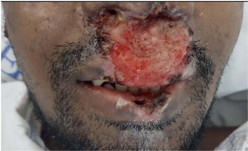
O avanço dos métodos diagnósticos, principalmente da biologia molecular, tornou a identificação dos microrganismos mais fácil e sensível. No entanto, esse avanço também exige uma melhor avaliação sobre o significado clínico do microrganismo isolado, uma vez que, em muitas situações, esse agente pode ser apenas um contaminante ou uma colonização. As micobactérias não tuberculosas (MNT) são bons exemplos desta dualidade e podem tanto causar uma gama de infecções clínicas quanto apenas colonizar determinados sítios do organismo. Neste relato, descrevemos o caso de um paciente previamente hígido com lesões ulceradas de pele e mucosas de evolução crônica e progressiva. Após diversos exames e avaliações, chegou-se ao diagnóstico de granulomatose com poliangiite. Todavia a biópsia da lesão nasolabial também identificou, por meio de biologia molecular, a presença de Mycobacterium mucogenicum. Essa identificação suscitou muitas dúvidas nas equipes médicas sobre o real papel desta MNT na patogênese ou no agravamento das lesões, com implicações decisórias sobre o seu tratamento (ou não) específico, uma vez que o paciente seria submetido a terapia imunossupressora.
Downloads
Referências
1. Band JD, Ward JI, Fraser DW, Peterson NJ, Silcox VA, Good RC, et al. Peritonitis due to a mycobacterium chelonei-like organism associated with intermittent chronic peritoneal dialysis. J Infect Dis. 1982 Jan;145(1):9-17. DOI: 10.1093/infdis/145.1.9
2. Esteban J, Martín-de-Hijas NZ, Fernandez AI, Fernandez-Roblas R, Gadea I; Madrid Study Group of Mycobacteria. Epidemiology of infections due to nonpigmented rapidly growing mycobacteria diagnosed in an urban area. Eur J Clin Microbiol Infect Dis. 2008 May;27(10):951-7. DOI: 10.1007/s10096-008-0521-7
3. Livni G, Yaniv I, Samra Z, Kaufman L, Solter E, Ashkenazi S, et al. Outbreak of Mycobacterium mucogenicum bacteraemia due to contaminated water supply in a paediatric haematology-oncology department. J Hosp Infect. 2008 Nov;70(3):253-8. DOI: 10.1016/j.jhin.2008.07.016
4. Adékambi T. Mycobacterium mucogenicum group infections: a review. Clin Microbiol Infect. 2009 Oct;15(10):911-8. DOI: 10.1111/j.1469-0691.2009.03028.x
5. Simões LC, Simões M, Vieira MJ. Biofilm interactions between distinct bacterial genera isolated from drinking water. Appl Environ Microbiol. 2007 Oct;73(19):6192-200. DOI: 10.1128/AEM.00837-07
6. Han XY, Dé I, Jacobson KL. Rapidly growing mycobacteria: clinical and microbiologic studies of 115 cases. Am J Clin Pathol. 2007 Jan;128(4):612-21. DOI: 10.1309/1KB2GKYT1BUEYLB5
7. Hawkins C, Qi C, Warren J, Stosor V. Catheter-related bloodstream infections caused by rapidly growing nontuberculous mycobacteria: a case series including rare species. Diagn Microbiol Infect Dis. 2008 Jun;61(2):187-91. DOI: 10.1016/j.diagmicrobio.2008.01.004
8. Gaviria JM, Garcia PJ, Garrido SM, Corey L, Boeckh M. Nontuberculous mycobacterial infections in hematopoietic stem cell transplant recipients: characteristics of respiratory and catheter-related infections. Biol Blood Marrow Transplant. 2000;6(4):361-9. DOI: 10.1016/s1083-8791(00)70012-7
9. Kline S, Cameron S, Streifel A, Yakrus MA, Kairis F, Peacock K, et al. An outbreak of bacteremias associated with Mycobacterium mucogenicum in a hospital water supply. Infect Control Hosp Epidemiol. 2004;25(12):1042-9. DOI: 10.1086/502341
10. Behra PRK, Pettersson BMF, Ramesh M, Dasgupta S, Kirsebom LA. Insight into the biology of Mycobacterium mucogenicum and Mycobacterium neoaurum clade members. Sci Rep. 2019 Dec;9(1):19259. DOI: 10.1038/s41598-019-55464-5
11. Redelman-Sidi G, Sepkowitz KA. Rapidly growing mycobacteria infection in patients with cancer. Clin Infect Dis. 2010 Aug;51(4):422-34. DOI: 10.1086/655140
12. Kodana M, Tarumoto N, Kawamura T, Saito T, Ohno H, Maesaki S, et al. Utility of the MALDI-TOF MS method to identify nontuberculous mycobacteria. J Infect Chemother. 2016 Jan;22(1):32-5. DOI: 10.1016/j.jiac.2015.09.006
13. Griffith DE, Aksamit T, Brown-Elliott BA, Catanzaro A, Daley C, Gordin F, et al; ATS Mycobacterial Diseases Subcommittee; American Thoracic Society; Infectious Disease Society of America. An official ATS/IDSA statement: diagnosis, treatment, and prevention of nontuberculous mycobacterial diseases. Am J Respir Crit Care Med. 2007;175(4):367-416. DOI: 10.1164/rccm.200604-571ST
14. Maz M, Chung SA, Abril A, Langford CA, Gorelik M, Guyatt G, et al. 2021 American College of Rheumatology/Vasculitis Foundation guideline for the management of giant cell arteritis and Takayasu arteritis. Arthritis Rheumatol. 2021 Jul;73(8):1349-65. DOI: 10.1002/art.41774
Downloads
Publicado
Como Citar
Edição
Seção
Licença
Copyright (c) 2023 Infectologia em Evidência

Este trabalho está licenciado sob uma licença Creative Commons Attribution 4.0 International License.
Todos os usuários podem ler, baixar, compartilhar e adaptar esta produção científica livremente para quaisquer fins (mesmo que comerciais), desde que seja dado o devido crédito aos autores e à publicação original e que qualquer alteração seja devidamente indicada.
> Ética
Todos os artigos publicados na Revista gozam de uma aprovação ética do Sistema Nacional de Ética em Pesquisa (antigo sistema CEP/CONEP) com base na Lei Federal 14.874/24 e outras regulamentações específicas brasileiras ou documento semelhante atestando a ciência e autorização por parte da instituição de origem no caso de trabalhos estrangeiros.
Os autores declaram não haver nenhum tipo de patrocínio ou conflito de interesses, salvo indicação em contrário no corpo do artigo.
Vale ressaltar que os relatos de caso são um valioso recurso de aprendizado para a comunidade científica, mas não devem ser utilizados isoladamente para guiar opções diagnósticas ou terapêuticas na prática clínica ou em políticas de saúde.


