
Piomiosite tuberculosa
DOI:
https://doi.org/10.5935/2764-734X.e20240442Palavras-chave:
Tuberculose extrapulmonar, Piomiosite, Abscesso, Diabetes mellitus, Relato de casoResumo
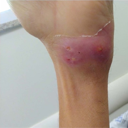
A tuberculose musculoesquelética representa uma rara forma de manifestação extrapulmonar da doença com poucos relatos na literatura médica, o que reforça a dificuldade no seu diagnóstico e o manejo em tempo adequado. Relatamos o caso de um paciente com quadro de piomiosite tuberculosa em múltiplos grupos musculares que apresentava como único antecedente patológico diabetes mellitus tipo II controlado. O diagnóstico foi estabelecido através da realização de teste rápido molecular (GeneXpert Mtbc/RIF®) em material purulento coletado por punção aspirativa de coleção dorsal. O paciente foi submetido a tratamento medicamentoso para tuberculose com esquema de rifampicina, isoniazida, pirazinamida e etambutol (RIPE) por dois meses em fase intensiva, seguido de manutenção com rifampicina e isoniazida (RI) por mais 10 meses, com resolução completa das coleções.
Downloads
Referências
1. Al-Khazraji A, Takher J, Alkhawam H, Fabbri M. Primary Tuberculous Pyomyositis of the Calf Muscles. Am J Med Sci. 2017;353(2):187-8. DOI: 10.1016/j.amjms.2016.05.010
2. Narayanappa G, Nandeesh BN. Infective myositis. Brain Pathol. 2021;31(3):e12950. DOI: 10.1111/bpa.12950
3. Puttick MP, Stein HB, Chan RM, Elwood RK, How AR, Reid GD. Soft tissue tuberculosis: a series of 11 cases. J Rheumatol [Internet]. 1995; [cited 20 Feb 2024]; 22(7):1321-5. Available from: https://pubmed.ncbi.nlm.nih.gov/7562766/
4. Murugesh AS, Edwin FM, Srinivasaprasad ND, Sujit S, Thirumalvalavan K. Tuberculous myositis and cellulitis in a renal transplant recipient. Indian J Tuberc. 2020;67(3):353-6. DOI: 10.1016/j.ijtb.2019.04.010
5. Rafaela S, Esther R, Carmen R, Marta S. MRI of musculoskeletal extraspinal tuberculosis. J Comput Assist Tomogr. 2001;25(2):177-83. DOI: 10.1097/00004728-200103000-00004
6. Simopoulou T, Varna A, Dailiana Z, Katsiari C, Alexiou I, Basdekis G, et al. Tuberculous pyomyositis: a re-emerging entity of many faces. Clin Rheumatol. 2016;35(4):1105-10. DOI: 10.1007/s10067-014-2564-8
7. Wang JY, Lee LN, Hsueh PR, Shih JY, Chang YL, Yang PC, et al. Tuberculous myositis: a rare but existing clinical entity. Rheumatology (Oxford). 2003;42(7):836-40. DOI: 10.1093/rheumatology/keg228
8. Habeych ME, Trinh T, Crum-Cianflone NF. Purulent infectious myositis (formerly tropical pyomyositis). J Neurol Sci. 2020:413:116767. DOI: 10.1016/j.jns.2020.116767
9. Narang S. Tuberculous pyomyositis of forearm muscles. Hand (NY). 2009;4(1):88-91. DOI: 10.1007/s11552-008-9127-x
10. Abdelwahab IF, Bianchi S, Martinoli C, Klein M, Hermann G. Atypical extraspinal musculoskeletal tuberculosis in immunocompetent patients: part II, tuberculous myositis, tuberculous bursitis, and tuberculous tenosynovites. Can Assoc Radiol J [Internet]. 2006; [cited 20 Feb 2024]; 57(5):278-86. Available from: https://pubmed.ncbi.nlm.nih.gov/17265982/
11. Thammaroj P, Panitchote A, Muktabhant C, Chowchuen P. Discrimination between tuberculous and bacterial pyomyositis in magnetic resonance features. Eur J Radiol Open. 2020:7:100214. DOI: 10.1016/j.ejro.2020.01.003
12. Ajantha GS, Shetty PC, Kulkarni RD, Biradar U. PCR as a diagnostic tool for extra-pulmonary tuberculosis. J Clin Diagn Res. 2013;7(6):1012-5. DOI: 10.7860/JCDR/2013/5425.3075
13. Lai YF, Chao TY, Wang YH, Lin AS. Pigtail drainage in the treatment of tuberculous pleural effusions: a randomised study. Thorax. 2003;58(2):149-51. DOI: 10.1136/thorax.58.2.149
Downloads
Publicado
Como Citar
Edição
Seção
Licença
Copyright (c) 2025 Infectologia em Evidência

Este trabalho está licenciado sob uma licença Creative Commons Attribution 4.0 International License.
Todos os usuários podem ler, baixar, compartilhar e adaptar esta produção científica livremente para quaisquer fins (mesmo que comerciais), desde que seja dado o devido crédito aos autores e à publicação original e que qualquer alteração seja devidamente indicada.
> Ética
Todos os artigos publicados na Revista gozam de uma aprovação ética do Sistema Nacional de Ética em Pesquisa (antigo sistema CEP/CONEP) com base na Lei Federal 14.874/24 e outras regulamentações específicas brasileiras ou documento semelhante atestando a ciência e autorização por parte da instituição de origem no caso de trabalhos estrangeiros.
Os autores declaram não haver nenhum tipo de patrocínio ou conflito de interesses, salvo indicação em contrário no corpo do artigo.
Vale ressaltar que os relatos de caso são um valioso recurso de aprendizado para a comunidade científica, mas não devem ser utilizados isoladamente para guiar opções diagnósticas ou terapêuticas na prática clínica ou em políticas de saúde.


