
Amputação de falanges como efeito adverso da injeção de penicilina benzatina
DOI:
https://doi.org/10.5935/2764-734X.e20240844Palavras-chave:
Síndrome de Nicolau, Penicilina G Benzatina, Amputados, Fasciotomia, Síndromes Compartimentais, Relato de casoResumo
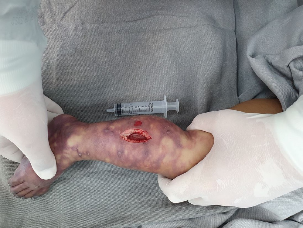
A Síndrome de Nicolau é um efeito adverso raro proveniente do uso de algumas medicações intramusculares. Relatamos aqui a evolução de uma criança de 13 meses atendida em pronto socorro depois de ter sido medicada há menos de 24 horas com penicilina benzatina devido a uma faringite. A criança desenvolveu uma isquemia grave do membro inferior homolateral em relação à região glútea onde o medicamento foi aplicado e, apesar de uma fasciotomia precoce visando mitigar uma síndrome compartimental, evoluiu com mumificação e amputação espontânea de falanges. A síndrome de Nicolau desponta como uma complicação possível da administração de antibióticos por via intramuscular e, em virtude de sua elevada morbidade, deve ser diagnosticada e tratada precocemente.
Downloads
Referências
1. Montané E, Santesmases J. Adverse drug reactions. Med Clin (Barc). 2020;154(5):178-84. DOI: 10.1016/j.medcli.2019.08.007
2. Kim SK, Kim TH, Lee KC. Nicolau syndrome after intramuscular injection: 3 cases. Arch Plast Surg. 2012;39(3):249-52. DOI: 10.5999/aps.2012.39.3.249
3. Corazza M, Capozzi O, Virgilit A. Five cases of livedo-like dermatitis (Nicolau’s syndrome) due to bismuth salts and various other non-steroidal anti-inflammatory drugs. J Eur Acad Dermatol Venereol. 2001;15(6):585-8. DOI: 10.1046/j.1468-3083.2001.00320.x
4. Cherasse A, Kahn MF, Mistrih R, Maillard H, Strauss J, Tavernier C. Nicolau’s syndrome after local glucocorticoid injection. Joint Bone Spine. 2003;70(5):390-2. DOI: 10.1016/s1297-319x(03)00137-4
5. Lardelli PF, Jermini LMM, Milani GP, Peeters GGAM, Ramelli GP, Zgraggen L, et al. Nicolau syndrome caused by non-steroidal anti-inflammatory drugs: Systematic literature review. Int J Clin Pract. 2020;74(10):e13567. DOI: 10.1111/ijcp.13567
6. Nagore E, Torrelo A, González-Mediero I, Zambrano A. Livedoid skin necrosis (Nicolau syndrome) due to triple vaccine (DTP) injection. Br J Dermatol. 1997;137(6):1030-1. DOI: 10.1111/j.1365-2133.1997.tb01585.x
7. Andre P, Haneke E. Nicolau syndrome due to hyaluronic acid injections. J Cosmet Laser Ther. 2016;18(4):239-44. DOI: 10.3109/14764172.2016.1157260
8. Faucher L, Marcoux D. What syndrome is this? Nicolau syndrome. Pediatr Dermatol. 1995;12(2):187-90. DOI: 10.1111/j.1525-1470.1995.tb00151.x
9. World Health Organization. World Health Organization model list of essential medicines: 21st list 2019 [Internet]. Geneva: WHO; 2018; [cited 2024 Aug 14] Available from: https://iris.who.int/handle/10665/325771
10. Kirby WM, Bulger RJ. The new penicillins and cephalosporins. Annu Rev Med. 1964:15:393-412. DOI: 10.1146/annurev.me.15.020164.002141
11. Green EA, Fogarty K, Ishmael FT. Penicillin Allergy: Mechanisms, Diagnosis, and Management. Prim Care. 2023;50(2):221-235. DOI: 10.1016/j.pop.2022.11.002
12. Haque MA, Nath ND, Johnston TV, Haruna S, Ahn J, Ovissipour R, et al. Harnessing biotechnology for penicillin production: Opportunities and environmental considerations. Sci Total Environ. 2024;946:174236. DOI: 10.1016/j.scitotenv.2024.174236
13. Atay S, Yilmaz Kurt F, Akkaya G, Karatağ G, Ilhan Demir Ş, Çalidağ U. Investigation of suitability of ventrogluteal site for intramuscular injections in children aged 36 months and under. J Spec Pediatr Nurs. 2017;22(4). DOI: 10.1111/jspn.12187
14. Chow TG, Ramsey AC. Penicillin Direct Challenges: Kids in the Lead, Adults Catching Up. J Allergy Clin Immunol Pract. 2024;12(2):458-9. DOI: 10.1016/j.jaip.2023.11.012
15. Yeniocak A, Kelahmetoğlu O, Özkan M, Temel M, Güneren E. A Basic Algorithmic Surgical Approach for Nicolau Syndrome. J Cutan Aesthet Surg. 2020;13(2):154-9. DOI: 10.4103/JCAS.JCAS_139_19
16. Gibson T. Karl Langer (1819-1887) and his lines. Br J Plast Surg. 1978;31(1):1-2. DOI: 10.1016/0007-1226(78)90002-4
17. Yazdani-Abyaneh MA, Griffith R, Falto-Aizpurua L, Nouri K. Famous lines in history: Langer lines. JAMA Dermatol. 2014;150(10):1087. DOI: 10.1001/jamadermatol.2014.659
18. Molho-Pessach V, Schaffer JV. Blaschko lines and other patterns of cutaneous mosaicism. Clin Dermatol. 2011;29(2):205-25. DOI: 10.1016/j.clindermatol.2010.09.012
19. Wu V 2nd, Tchanque-Fossuo CN, Stepenaskie S, Holguin T. Curvilinear violaceous plaques along Blaschko lines. JAAD Case Rep. 2021:12:29-31. DOI: 10.1016/j.jdcr.2021.04.004
20. Bezerra AS, Andrade AAAJ, Polimanti AC, Fürst RVC, Criado PR, Corrêa JA. Livedoid Vasculopathy: diagnosis and treatment in pregnant women. J Vasc Bras. 2020:19:e20190093. DOI: 10.1590/1677-5449.190093
21. Lopes JRA, D’Agostino Dias M, Correa JA, Batalha MAB, Guerra LKD. Randomized controlled clinical trial evaluating the efficacy of hyperbaric oxygen therapy in facilitating the healing of chronic foot ulcers in diabetic patients: the study protocol. Trials. 2020;21(1):816. DOI: 10.1186/s13063-020-04757-6
22. Lopes L, Filipe P, Alves A, Guerreiro F, Pires S. Nicolau syndrome after benzathine penicillin treated with hyperbaric oxygen therapy. Int J Dermatol. 2015;54(4):e103-6. DOI: 10.1111/ijd.12751
Downloads
Publicado
Como Citar
Edição
Seção
Licença
Copyright (c) 2025 Infectologia em Evidência

Este trabalho está licenciado sob uma licença Creative Commons Attribution 4.0 International License.
Todos os usuários podem ler, baixar, compartilhar e adaptar esta produção científica livremente para quaisquer fins (mesmo que comerciais), desde que seja dado o devido crédito aos autores e à publicação original e que qualquer alteração seja devidamente indicada.
> Ética
Todos os artigos publicados na Revista gozam de uma aprovação ética do Sistema Nacional de Ética em Pesquisa (antigo sistema CEP/CONEP) com base na Lei Federal 14.874/24 e outras regulamentações específicas brasileiras ou documento semelhante atestando a ciência e autorização por parte da instituição de origem no caso de trabalhos estrangeiros.
Os autores declaram não haver nenhum tipo de patrocínio ou conflito de interesses, salvo indicação em contrário no corpo do artigo.
Vale ressaltar que os relatos de caso são um valioso recurso de aprendizado para a comunidade científica, mas não devem ser utilizados isoladamente para guiar opções diagnósticas ou terapêuticas na prática clínica ou em políticas de saúde.


