
Herpes zoster e poliarterite nodosa: desafios diagnósticos
DOI:
https://doi.org/10.5935/2764-734X.e202112004Palavras-chave:
Poliarterite nodosa, Vasculite sistêmica, Algoritmos, Infecção pelo vírus da varicela-zoster, Relato de CasoResumo
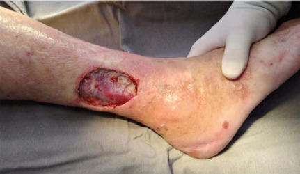
Paciente de 74 anos comparece ao hospital público universitário devido à dor em membros inferiores há 2 décadas, associado à lesão trófica em face lateral de perna direita há 3 meses e síndrome consumptiva há 2 meses. A paciente tratou herpes zoster em outro serviço médico por 4 anos. Após anamnese, exame físico e uso de algoritmo específico de vasculite primária, a paciente foi diagnosticada com poliarterite nodosa, permitindo início da terapêutica adequada e melhora clínica. Morfologicamente, o estudo histológico mostrou inflamação e necrose transmural em vasos de pequeno e médio calibre. O relato de caso evidencia a necessidade de uma investigação etiológica mais eficiente nos pacientes com vasculite primária.
Downloads
Referências
1. Gnann Junior JW, Whitley RJ. Clinical practice. Herpes zoster. N Engl J Med. 2002 Aug;347(5):340-6.
2. Rosamilia LL. Herpes zoster presentation, management, and prevention: a modern case-based review. Am J Clin Dermatol. 2020 Feb;21(1):97-107.
3. Ehrenstein B. Diagnosis, treatment and prophylaxis of herpes zoster. Z Rheumatol. 2020 Dec;79(10):1009-17.
4. Callaghan BC, Price RS, Feldman EL. Distal symmetric polyneuropathy: a review. JAMA. 2015 Nov;314(20):2172-81.
5. Lau CH, Missotten T, Salzmann J, Lightman SL. Acute retinal necrosis features, management, and outcomes. Ophthalmology. 2007 Apr;114(4):756-62.
6. Hanewinckel R, Drenthen J, Van Oijen M, Hofman A, Van Doorn PA, Ikram MA. Prevalence of polyneuropathy in the general middle-aged and elderly population. Neurology. 2016 Nov;87(18):1892-8.
7. Jennette JC, Falk RJ, Bacon PA, Basu N, Cid MC, Ferrario F, et al. 2012 revised International Chapel Hill Consensus Conference nomenclature of vasculitides. Arthritis Rheum. 2013 Jan;65(1):1-11.
8. Criado PR, Marques GF, Morita TC, Carvalho JF. Epidemiological, clinical and laboratory profiles of cutaneous polyarteritis nodosa patients: report of 22 cases and literature review. Autoimmun Rev. 2016 Jun;15(6):558-63.
9. Bezerra AS, Polimanti AC, Oliveira RA, Fürst RVC, Criado PR, Corrêa JA. Early diagnosis and treatment of leukocytoclastic vasculitis: case report. J Vasc Bras. 2020 Jan;19:e20180072.
10. Jennette JC, Falk RJ, Andrassy K, Bacon PA, Churg J, Gross WL, et al. Nomenclature of systemic vasculitides. Proposal of an international consensus conference. Arthritis Rheum. 1994 Feb;37(2): 187-92.
11. Ozen S. The changing face of polyarteritis nodosa and necrotizing vasculitis. Nat Rev Rheumatol. 2017 Jun;13(6):381-6.
12. Pagnoux C, Seror R, Henegar C, Mahr A, Cohen P, Le Guern V, et al. Clinical features and outcomes in 348 patients with polyarteritis nodosa: a systematic retrospective study of patients diagnosed between 1963 and 2005 and entered into the French Vasculitis Study Group Database. Arthritis Rheum. 2010 Feb;62(2):616-26.
13. Puéchal X, Pagnoux C, Baron G, Quémeneur T, Néel A, Agard C, et al. Adding azathioprine to remission-induction glucocorticoids for eosinophilic granulomatosis with polyangiitis (Churg-Strauss), microscopic polyangiitis, or polyarteritis nodosa without poor prognosis factors: a randomized, controlled trial. Arthritis Rheumatol. 2017 Nov;69(11):2175-86.
14. Bezerra AS, Polimanti AC, Fürst RVC, Corrêa JA. Algorithm for diagnosis of primary vasculitides. J Vasc Bras. 2019 Mar;18:e20180092.
15. Azanza JC, Sarmiento PC, Lia NL, Alexander AS, Modi V. Leukocytoclastic vasculitis: an early skin biopsy makes a difference. Cureus. 2020 May;12(5):e7912.
16. Lightfoot Junior RW, Michel BA, Bloch DA, Hunder GG, Zvaifler NJ, McShane DJ, et al. The American College of Rheumatology 1990 criteria for the classification of polyarteritis nodosa. Arthritis Rheum. 1990 Aug;33(8):1088-93.
17. Ribi C, Cohen P, Pagnoux C, Mahr A, Arène JP, Puéchal X, et al. Treatment of polyarteritis nodosa and microscopic polyangiitis without poor-prognosis factors: a prospective randomized study of one hundred twenty-four patients. Arthritis Rheum. 2010 Apr;62(4):1186-97.
18. Ushiyama S, Shimojima Y, Ueno KI, Kishida D, Miyazaki D, Sekijima Y. Clinical characteristics of patients with myalgia as the initial manifestation of small and medium-sized vasculitis: a retrospective study. Rheumatol Int. 2020;40:1667-74.
19. Martins-Martinho J, Dourado E, Khmelinskii N, Espinosa P, Ponte C. Localized forms of vasculitis. Curr Rheumatol Rep. 2021 Jul;23(7):49.
20. Krusche M, Ruffer N, Kötter I. Tocilizumab treatment in refractory polyarteritis nodosa: a case report and review of the literature. Rheumatol Int. 2019 Feb;39(2):337-44.
Downloads
Publicado
Como Citar
Edição
Seção
Licença
Copyright (c) 2022 Infectologia em Evidência

Este trabalho está licenciado sob uma licença Creative Commons Attribution 4.0 International License.
Todos os usuários podem ler, baixar, compartilhar e adaptar esta produção científica livremente para quaisquer fins (mesmo que comerciais), desde que seja dado o devido crédito aos autores e à publicação original e que qualquer alteração seja devidamente indicada.
> Ética
Todos os artigos publicados na Revista gozam de uma aprovação ética do Sistema Nacional de Ética em Pesquisa (antigo sistema CEP/CONEP) com base na Lei Federal 14.874/24 e outras regulamentações específicas brasileiras ou documento semelhante atestando a ciência e autorização por parte da instituição de origem no caso de trabalhos estrangeiros.
Os autores declaram não haver nenhum tipo de patrocínio ou conflito de interesses, salvo indicação em contrário no corpo do artigo.
Vale ressaltar que os relatos de caso são um valioso recurso de aprendizado para a comunidade científica, mas não devem ser utilizados isoladamente para guiar opções diagnósticas ou terapêuticas na prática clínica ou em políticas de saúde.


