
Purpura fulminans na doença meningocócica invasiva
DOI:
https://doi.org/10.5935/2764-734X.e202201006Palavras-chave:
Neisseria meningitidis, Púrpura Fulminante, Infecções Meningocócicas, Neisseria meningitidis Sorogrupo W-135Resumo
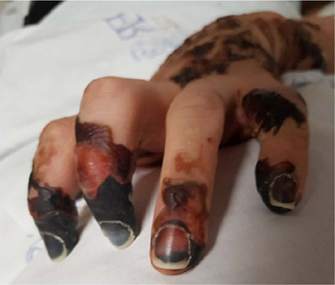
A doença meningocócica já foi responsável por milhões de mortes em suas epidemias no mundo todo. O objetivo deste relato é destacar a rápida evolução dessa doença atualmente menos prevalente e sua principal complicação, purpura fulminans, uma síndrome rara e altamente letal, assim como comentar seu manejo clínico e cirúrgico. Trata-se de uma paciente do sexo feminino de 23 anos, profissional da saúde, que foi diagnosticada com meningoccemia por N. meningitidis sorogrupo W, cursando clinicamente com purpura fulminans e posterior necessidade de amputação dos segmentos acometidos, além de outras complicações. A paciente em questão não era vacinada para este sorogrupo, o que teria evitado este quadro grave. Há de se considerar a vacinação para o grupo de risco ao qual ela pertencia. Este relato é importante para não nos esquecermos das doenças que há décadas ou séculos atrás geraram grandes problemas sanitários e atualmente estão controladas.
Downloads
Referências
1. Focaccia R. Doença meningocócica. In: Veronesi R, Focaccia R, eds. Tratado de infectologia. 5ª ed. São Paulo: Atheneu; 2015. p. 1053-66.
2. Ministério da Saúde (BR). Secretaria de Vigilância em Saúde. Situação epidemiológica da doença meningocócica, Brasil, 2007-2013. Boletim epidemiológico - volume 47, nº 29, 2016 [Internet]. Brasília (DF): Ministério da Saúde; 2020; [acesso em 2020 Dez 20]. Disponível em: https://www.gov.br/saude/pt-br/assuntos/boletins-epidemiologicos/numeros-anteriores
3. Ministério da Saúde (BR). Meningite [Internet]. Brasília (DF): Ministério da Saúde; 2020; [acesso em 2020 Dez 20]. Disponível em: https://www.gov.br/saude/pt-br/assuntos/saude-de-a-a-z/m/meningite
4. Guiddir T, Gros M, Hong E, Terrade A, Denizon M, Deghmane AE, et al. Unusual initial abdominal presentations of invasive meningococcal disease. Clin Infect Dis. 2018 Oct;67(8):1220-7. DOI: https://doi.org/10.1093/cid/ciy257
5. Evans L, Rhodes A, Alhazzani W, Antonelli M, Coopersmith CM, French C, et al. Surviving sepsis campaign: international guidelines for management of sepsis and septic shock 2021. Crit Care Med. 2021 Nov;49(11):e1063-e43. DOI: https://doi.org/10.1097/CCM.0000000000005337
6. Abe R, Oda S, Sadahiro T, Nakamura M, Hirayama Y, Tateishi Y, et al. Gram-negative bacteremia induces greater magnitude of inflammatory response than Gram-positive bacteremia. Crit Care. 2010 Mar;14(2):R27. DOI: https://doi.org/10.1186/cc8898
7. Chalmers E, Cooper P, Forman K, Grimley C, Khair K, Minford A, et al. Purpura fulminans: recognition, diagnosis and management. Arch Dis Child. 2011;96(11):1066-71. DOI: https://doi.org/10.1136/adc.2010.199919
8. Colling ME, Bendapudi PK. Purpura fulminans: mechanism and management of dysregulated hemostasis. Transfus Med Rev. 2018 Apr;32(2):69-76. DOI: https://doi.org/10.1016/j.tmrv.2017.10.001
9. Ministério da Saúde (BR). Secretaria de Vigilância em Saúde. Departamento de Imunização e Doenças Transmissíveis. Coordenação-Geral do Programa Nacional de Imunizações. Manual dos centros de referência para imunobiológicos especiais [Internet]. 5ª ed. Brasília (DF): Ministério da Saúde; 2019; [acesso em 2020 Dez 20]. Disponível em: https://pesquisa.bvsalud.org/bvsms/resource/pt/mis-40552
Downloads
Publicado
Como Citar
Edição
Seção
Licença
Copyright (c) 2022 Infectologia em Evidência

Este trabalho está licenciado sob uma licença Creative Commons Attribution 4.0 International License.
Todos os usuários podem ler, baixar, compartilhar e adaptar esta produção científica livremente para quaisquer fins (mesmo que comerciais), desde que seja dado o devido crédito aos autores e à publicação original e que qualquer alteração seja devidamente indicada.
> Ética
Todos os artigos publicados na Revista gozam de uma aprovação ética do Sistema Nacional de Ética em Pesquisa (antigo sistema CEP/CONEP) com base na Lei Federal 14.874/24 e outras regulamentações específicas brasileiras ou documento semelhante atestando a ciência e autorização por parte da instituição de origem no caso de trabalhos estrangeiros.
Os autores declaram não haver nenhum tipo de patrocínio ou conflito de interesses, salvo indicação em contrário no corpo do artigo.
Vale ressaltar que os relatos de caso são um valioso recurso de aprendizado para a comunidade científica, mas não devem ser utilizados isoladamente para guiar opções diagnósticas ou terapêuticas na prática clínica ou em políticas de saúde.


