
Doença de Castleman multicêntrica em paciente infectado pelo vírus da imunodeficiência humana
DOI:
https://doi.org/10.5935/2764-734X.e202205010Palavras-chave:
Hiperplasia do Linfonodo Gigante, Linfoma Relacionado a AIDS, Herpesvirus Humano 8, Síndrome Inflamatória da Reconstituição Imune, Sarcoma de KaposiResumo
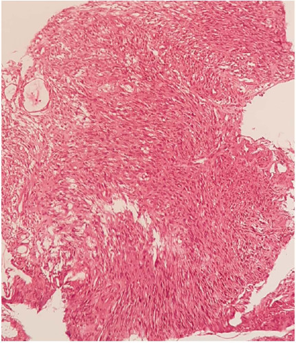
Doença de Castleman consiste num distúrbio linfoproliferativo raro com apresentações clínicas, histológicas e etiológicas distintas, podendo se apresentar como linfadenopatia localizada ou doença sistêmica. A apresentação da Doença de Castleman multicêntrica tem associação com herpes vírus humano-8 sobretudo em pacientes infectados pelo vírus da imunodeficiência humana e ocasionalmente é encontrado com Sarcoma de Kaposi. Apresentamos um caso pouco descrito na literatura de um homem de 41 anos com diagnóstico recente de infecção pelo vírus da imunodeficiência humana e elevada carga viral que evoluiu com linfonodomegalia periférica associada a Doença de Castleman multicêntrica e Sarcoma de Kaposi após o início de terapia antirretroviral.
Downloads
Referências
1. Cabot RC, Founder BC, Town VW. Case records of the Massachusetts general hospital weekly clinicopathological exercises: case 40011. N Engl J Med. 1954 Jan;250(1):26-30. DOI: https://www.nejm.org/doi/full/10.1056/NEJM195401072500107
2. Haq IU, Pria AD, Papanastasopoulos P, Stegmann K, Bradshaw D, Nelson M, et al. The clinical application of plasma Kaposi sarcoma herpesvirus viral load as a tumour biomarker: results from 704 patients. HIV Med. 2016 Jan;17(1):56-61.
3. Mylona EE, Baraboutis IG, Lekakis LJ, Ourania G, Papastamopoulos VV, Skoutelis A. Multicentric Castleman’s disease in HIV infection: a systematic review of the literature. AIDS Rev. 2008 Jan/Mar;10(1):25-35.
4. Oksenhendler E, Boutboul D, Fajgenbaum D, Mirouse A, Fieschi C, Malphettes M, et al. The full spectrum of Castleman disease: 273 patients studied over 20 years. Br J Haematol. 2018 Jan;180(2):206-16.
5. Uldrick TS, Wang V, O’Mahony D, Aleman K, Wyvill KM, Marshall V, et al. An interleukin-6-related systemic inflammatory syndrome in patients co-infected with Kaposi sarcoma-associated herpesvirus and HIV but without Multicentric Castleman disease. Clin Infect Dis. 2010 Ago;51(3):350-8.
6. Lachant NA, Sun NC, Leong LA, Oseas RS, Prince HE. Multicentric angiofollicular lymph node hyperplasia (Castleman’s disease) followed by Kaposi’s sarcoma in two homosexual males with the acquired immunodeficiency syndrome (AIDS). Am J Clin Pathol. 1985 Jan;83(1):27-33.
7. Aoki Y, Tosato G, Fonville TW, Pittaluga S. Serum viral interleukin-6 in AIDS-related multicentric Castleman disease. Blood. 2001 Abr;97(8):2526-7.
8. Bower M, Newsom-Davis T, Naresh K, Merchant S, Lee B, Gazzard B, et al. Clinical features and outcome in HIV-associated multicentric Castleman’s disease. J Clin Oncol. 2011 Jun;29(18):2481-6.
9. Aaron L, Lidove O, Yousry C, Roudiere L, Dupont B, Viard J. Human herpesvirus 8-positive Castleman disease in human immunodeficiency vírus-infected patients: the impact of highly active antiretroviral therapy. Clin Infect Dis. 2002 Out;35(7):880-2.
10. Zietz C, Bognor JR, Goebel FD, Löhrs U. An unusual cluster of cases of Castleman’s disease during highly active antiretroviral therapy for AIDS. N Engl J Med. 1999 Jun;340(24):1923-4.
11. Lee SM, Edwards SG, Chilton DN, Ramsay A, Miller RF. Highly active antiretroviral therapy alone may be an effective treatment for HIV-associated multicentric Castleman’s disease. Haematologica. 2010;95(11):1979-81.
12. Powles T, Stebbing J, Bazeos A, Hatzimichael E, Mandalia S, Nelson M, et al. The role of immune suppression and HHV-8 in the increasing incidence of HIV-associated multicentric Castleman’s disease. Ann Oncol. 2009 Abr;20(4):775-9.
13. Oksenhendler E, Boulanger E, Galicier L, Ming-Qing D, Dupin N, Diss TC, et al. High incidence of Kaposi sarcoma-associated herpesvirus-related non-Hodgkin lymphoma in patients with HIV infection and multicentric Castleman disease. Blood. 2002 Abr;99(7):2331-6.
14. Bottieau E, Colebunders R, Schroyens W, Van Droogenbroeck J, Droogh E, Depraetere K, et al. Multicentric Castleman’s disease in 2 patients with HIV infection, unresponsive to antiviral therapy. Acta Clin. 2000;55:97-101.
15. Goncalves PH, Ziegelbauer J, Uldrick TS, Yarchoan R. Kaposi sarcoma herpesvirus-associated cancers and related diseases. Curr Opin HIV AIDS. 2017 Jan;12(1):47-56.
16. Guo WX, Antakly T, Cadotte M, Kachra Z, Kunkel L, Masood R, et al. Expression and cytokine regulation of glucocorticoid receptors in Kaposi’s sarcoma. Am J Pathol. 1996 Jun;148(6):1999-2008.
17. Polizzotto MN, Uldrick TS, Wyvill KM, Aleman K, Marshall V, Wang V, et al. Clinical features and outcomes of patients with symptomatic Kaposi Sarcoma Herpesvirus (KSHV)-associated inflammation: Prospective characterization of KSHV inflammatory cytokine syndrome (KICS). Clin Infect Dis. 2016 Mar;62(6):730-8.
18. Uldrick TS, Polizzotto MN, Aleman K, Wyvill KM, Marshall V, Whitby D, et al. Rituximab plus liposomal doxorubicin in HIV infected patients with KSHV-associated multicentric Castleman disease. Blood. 2014 Dez;124(24):3544-52.
19. Hofmann C, Schmid H, Müller M, Teutsch C, Van Lunzen J, Esser S, et al. Improved outcome with rituximab in patients with HIV-associated multicentric Castleman disease. Blood. 2011 Set;118(13):3499-503
Downloads
Publicado
Como Citar
Edição
Seção
Licença
Copyright (c) 2022 Infectologia em Evidência

Este trabalho está licenciado sob uma licença Creative Commons Attribution 4.0 International License.
Todos os usuários podem ler, baixar, compartilhar e adaptar esta produção científica livremente para quaisquer fins (mesmo que comerciais), desde que seja dado o devido crédito aos autores e à publicação original e que qualquer alteração seja devidamente indicada.
> Ética
Todos os artigos publicados na Revista gozam de uma aprovação ética do Sistema Nacional de Ética em Pesquisa (antigo sistema CEP/CONEP) com base na Lei Federal 14.874/24 e outras regulamentações específicas brasileiras ou documento semelhante atestando a ciência e autorização por parte da instituição de origem no caso de trabalhos estrangeiros.
Os autores declaram não haver nenhum tipo de patrocínio ou conflito de interesses, salvo indicação em contrário no corpo do artigo.
Vale ressaltar que os relatos de caso são um valioso recurso de aprendizado para a comunidade científica, mas não devem ser utilizados isoladamente para guiar opções diagnósticas ou terapêuticas na prática clínica ou em políticas de saúde.


