
Vimblastina intralesional em Sarcoma de Kaposi Oral
DOI:
https://doi.org/10.5935/2764-734X.e202201008Palavras-chave:
Sarcoma de Kaposi, Injeções, Intralesionais, Vimblastina, Herpesvirus Humano 8, HIVResumo
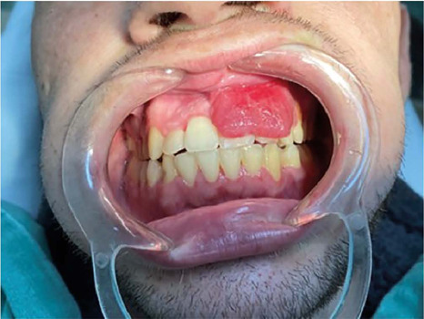
Sarcoma de Kaposi (SK) é um tumor multicêntrico, caracterizado por lesões avermelhadas ou violáceas em pele, mucosa bucal e outras localizações. Associado ao Herpesvírus tipo 8, é mais prevalente entre homens que fazem sexo com homens (HSH), e a boca pode ser o único local envolvido. Tratamentos para o SK oral incluem remoção cirúrgica, radioterapia e a quimioterapia intralesional, entre outros. O objetivo deste Relato de Caso foi descrever e analisar a eficácia do tratamento de SK oral em paciente com aids através de injeções locais de vimblastina, visando destacar seus resultados positivos num contexto de tratamento complementar da neoplasia.
Downloads
Referências
1. Karamanou M, Antoniou C, Stratigos AJ, Saridaki Z, Androutsos G. The eminent dermatologist Moriz Kaposi (1837-1902) and the first description of idiopathic multiple pigmented sarcoma of the skin. J BUON. 2013 Oct/Dec;18(4):1101-5.
2. Feit H. Sarcoma (Kaposi). Arch Derm Syphilol. 1928;18:611–2 apud Aroua A, Ali RB, Chokri A, Selmi J. Solitary Kaposi’s sarcoma in the oral cavity of an HIV-negative patient: a rare case report. Sch J Med Case Rep. 2017;5(11):713-6.
3. Dittmer DP, Damania B. Kaposi sarcoma-associated herpesvirus: immunobiology, oncogenesis, and therapy. J Clin Invest. 2016 Sep;126(9):3165-75.
4. Beral V, Peterman TA, Berkelman RL, Jaffe HW. Kaposi’s sarcoma among persons with AIDS: a sexually transmitted infection? Lancet. 1990 Jan;335(8682):123-8.
5. Epstein JB. Treatment of oral Kaposi sarcoma with intralesional vinblastine. Cancer. 1993 Mar;71(5):1722-5.
6. Epstein JB, Lozada-Nur F, McLeod WA, Spinelli J. Oral Kaposi’s sarcoma in acquired immunodeficiency syndrome. Review of management and report of the efficacy of intralesional vinblastine. Cancer. 1989 Dec;64(12):2424-30.
7. Fatahzadeh M, Schwartz RA. Oral Kaposi’s sarcoma: a review and update. Int J Dermatol. 2013 Jun;52(6):666-72.
8. McCormick SU. Intralesional vinblastine injections for the treatment of oral Kaposi’s sarcoma: report of 10 patients with 2-year follow-up. J Oral Maxillofac Surg. 1996 May;54(5):583-9.
9. Volberding P, Conant MA, Stricker RB, Lewis BJ. Chemotherapy in advanced Kaposi’s sarcoma. Implications for current cases in homosexual men. Am J Med. 1983 Apr;74(4):652-6.
10. Goncalves PH, Uldrick TS, Yarchoan R. HIV-associated Kaposi sarcoma and related diseases. AIDS. 2017 Sep;31(14):1903-16.
11. Chang Y, Cesarman E, Pessin MS, Lee F, Culpepper J, Knowles DM, et al. Identification of herpesvirus-like DNA sequences in AIDS-associated Kaposi’s sarcoma. Science. 1994 Dec;266(5192):1865-9.
12. Gorsky M, Epstein JB. A case series of acquired immunodeficiency syndrome patients with initial neoplastic diagnoses of intraoral Kaposi’s sarcoma. Oral Surg Oral Med Oral Pathol Oral Radiol Endod. 2000 Nov;90(5):612-7.
13. Lebbe C, Blum L, Pellet C, Blanchard G, Verola O, Morel P, et al. Clinical and biological impact of antiretroviral therapy with protease inhibitors on HIV-related Kaposi’s sarcoma. AIDS. 1998 May;12(7):F45-9.
Downloads
Publicado
Como Citar
Edição
Seção
Licença
Copyright (c) 2022 Infectologia em Evidência

Este trabalho está licenciado sob uma licença Creative Commons Attribution 4.0 International License.
Todos os usuários podem ler, baixar, compartilhar e adaptar esta produção científica livremente para quaisquer fins (mesmo que comerciais), desde que seja dado o devido crédito aos autores e à publicação original e que qualquer alteração seja devidamente indicada.
> Ética
Todos os artigos publicados na Revista gozam de uma aprovação ética do Sistema Nacional de Ética em Pesquisa (antigo sistema CEP/CONEP) com base na Lei Federal 14.874/24 e outras regulamentações específicas brasileiras ou documento semelhante atestando a ciência e autorização por parte da instituição de origem no caso de trabalhos estrangeiros.
Os autores declaram não haver nenhum tipo de patrocínio ou conflito de interesses, salvo indicação em contrário no corpo do artigo.
Vale ressaltar que os relatos de caso são um valioso recurso de aprendizado para a comunidade científica, mas não devem ser utilizados isoladamente para guiar opções diagnósticas ou terapêuticas na prática clínica ou em políticas de saúde.


